Tech
Evolving Organic Technology of the Human Eye

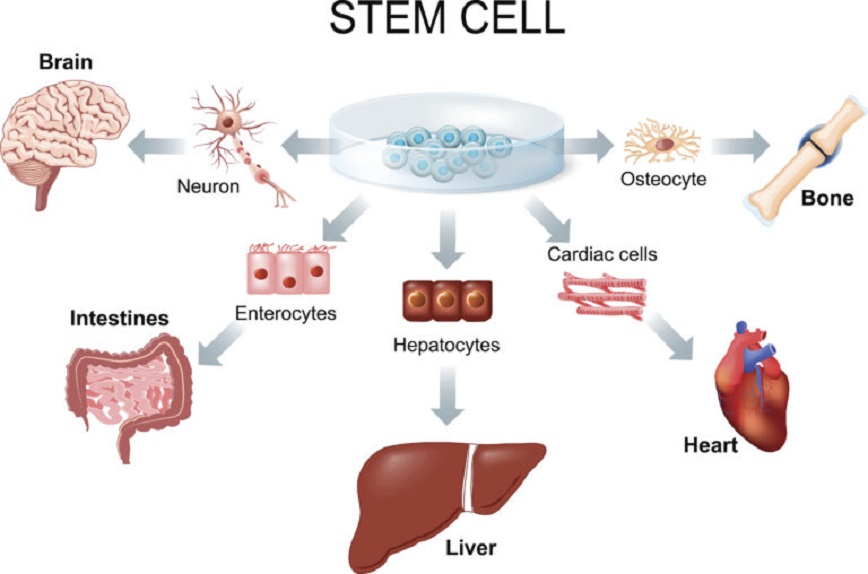
LAST MONTH, THERE WAS NEWS THAT RESEARCHERS AT THE INSTITUTE OF BASIC SCIENCES (IBS) HAD DEVELOPED A “BRAIN ORGANOID CULTURE PLATFORM” THAT IMPLEMENTED AN ENVIRONMENT SIMILAR TO THAT OF A REAL HUMAN BRAIN. ‘ORGANOID’ REFERS TO A ‘LONG-TERM ANALOGUE’ THAT INDUCES STEM CELLS TO A SPECIFIC BODY ORGAN AND REPRODUCES THEM SIMILARLY TO ORGANS IN VITRO. ORGANIC TECHNOLOGY HAS BEEN CONTINUOUSLY DEVELOPING FOR NEW DRUG DEVELOPMENT, DISEASE TREATMENT, AND IN-FACTORY DEVELOPMENT. IN 2019, THERE WERE REPORTS THAT IN THE CASE OF BRAIN ORGANOIDS, WAVELENGTHS CORRESPONDING TO THE PRE-CONSCIOUSNESS STAGE HAD REACHED THE LEVEL AT WHICH WAVELENGTHS WERE DETECTED BY CREATING A SO-CALLED “MINI-BRAIN” THAT WAS VERY SMALL TO BE CALLED A BRAIN. THE BRAIN ORGANOID PLATFORM IS NOT ONLY MATURE CLOSE TO THE NEWBORN BRAIN LEVEL BUT ALSO MORE THAN TWICE THE SIZE OF THE PREVIOUS ONE.
this time, the news is that the brain organoids have created snow. a recent experimental paper published in cell stem cells reported that after turning human stem cells into precursor stages of prefrontal neurons in our brain, these cells formed a round, hollow cup-shaped structure similar to two eyes. the organoids were also electrically engaged when exposed to light, according to the report.
the eye is an important body organ that we need to process light and visual information. the very complex and sophisticated process of eye development in our body has been studied so far, for example, during the 19th century it has been found that the retina develops and protrudes from the cerebral side of the frontal lobe.’
two studies published in 2011 and 2012 showed that retina can be induced to form in rat and human stem cells, respectively. however, in the process of embryo development, as eye vesicles develop in the cerebellum of the frontal lobe, the researchers in this published study wanted to reproduce this process using organoids rather than cells.

eye vesicles develop in brain organoids
first, the researchers who modified the existing protocol to induce brain organoids added retinic acid, which is essential for eye development. it induced the development of the eye in organoids, and in fact, after 30 days, the researchers observed the formation of a colored structure in the area corresponding to the eye. these eye structures were symmetrically two, which were identified in 86 of the 95 organoids, the researchers added.
And using immunostaining, a method of detecting proteins using antibodies, it was confirmed that there are proteins present in the precursor cells of the eye, such as RAX or FOXG1, in the eye area. RNA-seq analysis to identify genes transcribed with RNA before proteins were made also confirmed genes related to eye development and retinal regeneration.
in the snow parcels, the small organs peculiar to the eyes appear.
Subsequent brain organoids gradually developed eye vesicles, and eye-related cells such as the retina began to be observed. After 60 days, noticeable structures began to occur in the brain’s eye vesicles. Using immunofluorescence staining, the researchers confirmed the presence of organelles in our eyes, such as early-stage crystals and corneal cell layers, in the eye vesicle structure of organoids. It was also confirmed that there was a signaling reaction in the organoid by measuring the signaling of the retina and the ‘electroretinography’ which measures the signaling of the retina, or by exposing it to light stress with different intensities and measuring its response.
the study used brain organoids to develop two symmetrical round, hollow eyes, and experimentally confirmed that their cellular features and functions were similar to real eyes. however, it has not reproduced a very mature stage of the retina, and although technologies that can continuously develop and maintain organoids after 60 days shown in the study should be tried in the future, this study is being evaluated as a groundbreaking study that has shown the possibility of brain organoids being used as models for studying eye-related studies, especially retina-related diseases.